AI-Powered Precision: Enhancing CT and MRI Diagnoses

The Role of AI in CT and MRI Diagnosis:
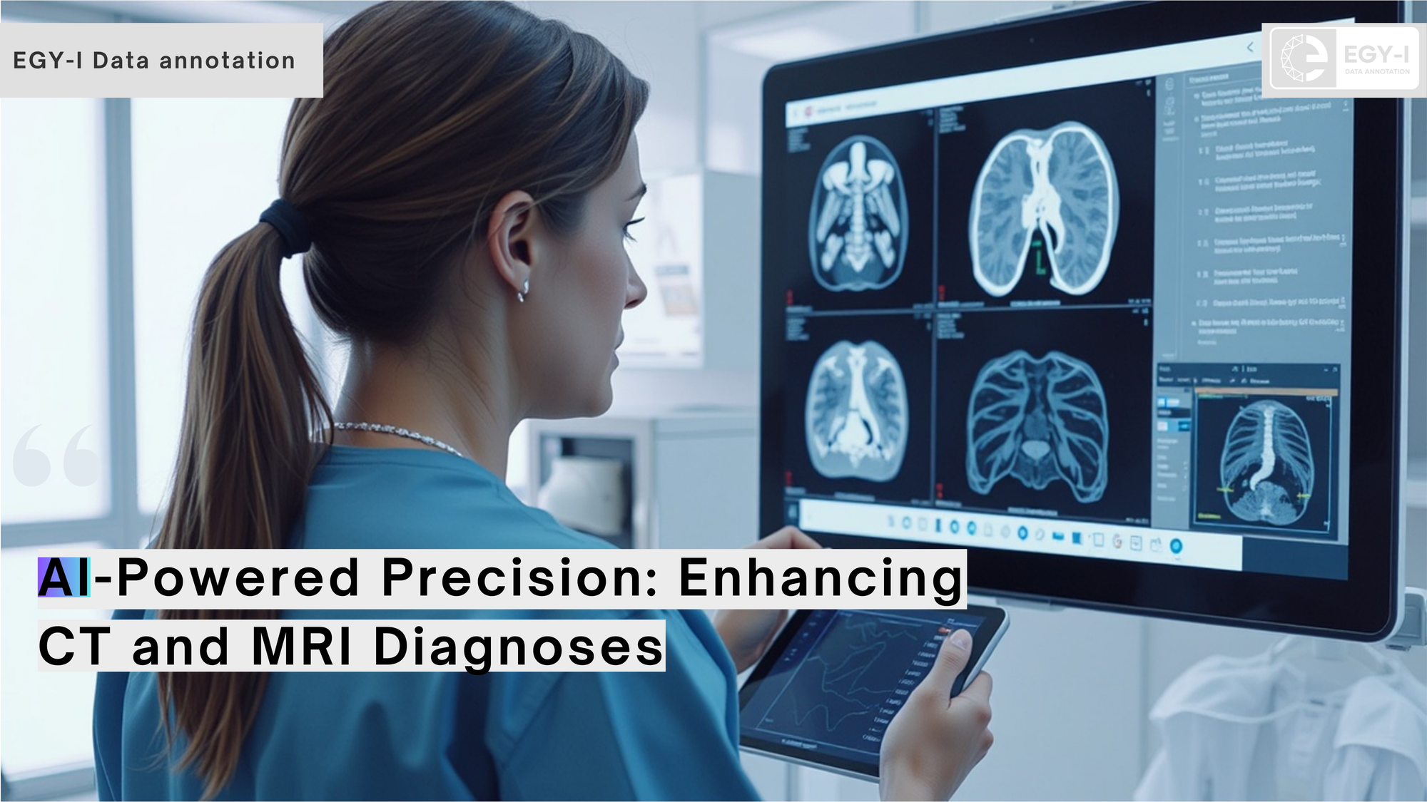
In the ever-evolving field of medical imaging, Artificial Intelligence (AI) is playing a transformative role, particularly in CT (Computed Tomography) and MRI (Magnetic Resonance Imaging) diagnostics. These imaging modalities are critical for detecting a wide range of medical conditions, from cancers and neurological disorders to trauma and cardiovascular diseases. However, the sheer volume and complexity of data generated by CT and MRI scans can overwhelm even the most experienced radiologists.
EGY-I, is at the forefront of enabling AI-driven solutions for CT and MRI diagnostics. Our high-quality annotated datasets empower AI developers to create tools that enhance diagnostic accuracy, optimize workflows, and improve patient outcomes.
Why CT and MRI Imaging Need AI?
CT and MRI are known for their ability to provide detailed and high-resolution images. Despite their advantages, these imaging technologies face significant challenges:
- Massive Data Volumes: A single CT or MRI scan can generate hundreds to thousands of images, requiring extensive analysis.
- Time-Intensive Process: Manual interpretation can delay diagnosis, especially in emergency cases.
- Risk of Human Error: Subtle abnormalities may go unnoticed due to fatigue or high workloads.
This is where AI in CT and MRI diagnostics proves invaluable. By automating tasks like abnormality detection and image segmentation, AI reduces the burden on radiologists and ensures faster, more accurate diagnoses.
Key Applications of AI in CT and MRI Diagnostics
1. Automated Abnormality DetectionAI algorithms are capable of identifying and classifying abnormalities with precision.
- Tumor Detection: AI helps in early detection of brain, liver, and lung tumors by analyzing subtle changes in tissue structure.
- Stroke Diagnosis: In brain CT and MRI scans, AI can differentiate between ischemic and hemorrhagic strokes, enabling timely intervention.
- Bone Fractures: CT scans of complex fractures are analyzed quickly and accurately by AI, assisting orthopedic specialists.

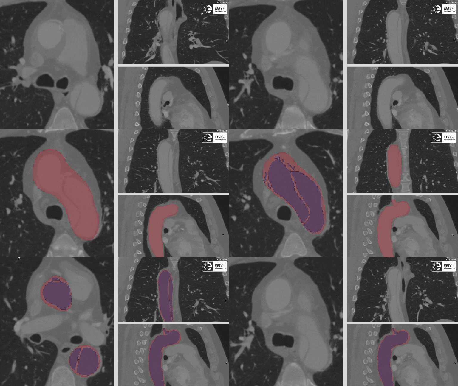
2. Advanced Image SegmentationAI excels in segmenting organs, tissues, and pathological regions, facilitating accurate diagnosis and treatment planning.
- Tumor Segmentation: Precise measurement of tumor size and shape for monitoring growth or treatment response.
- Organ Mapping: AI delineates organs such as the brain, heart, and liver to assist in radiation therapy and surgical planning.

3. Workflow OptimizationAI improves efficiency by streamlining radiology workflows.
- Case Prioritization: AI flags critical cases, ensuring life-threatening conditions are reviewed first.
- Preliminary Reports: AI generates initial findings, reducing radiologists’ workload and turnaround time.
4. Enhanced Image QualityAI enhances CT and MRI scans by:
- Reducing noise and correcting artifacts.
- Improving resolution, ensuring clearer and more accurate imaging for better diagnoses.
5. Functional and Specialized ImagingAI supports advanced imaging techniques such as:
- fMRI: Functional MRI uses AI to map brain activity for neurological studies.
- Dual-Energy CT: AI differentiates tissue types for enhanced diagnostic capabilities.
The Importance of Image Annotation for AI in CT and MRI
The success of AI in CT and MRI diagnostics hinges on high-quality annotated datasets. Annotation involves meticulously labeling images to highlight structures, abnormalities, or other points of interest.EGY-I specializes in providing world-class annotation services for CT and MRI datasets. Our annotations support the development of AI tools that are not only accurate but also clinically reliable.
Our Annotation Capabilities Include:
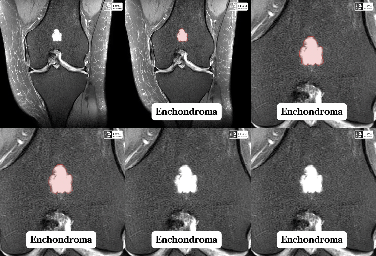
- Lesion Marking: Highlighting tumors, cysts, or other abnormalities in CT and MRI scans.
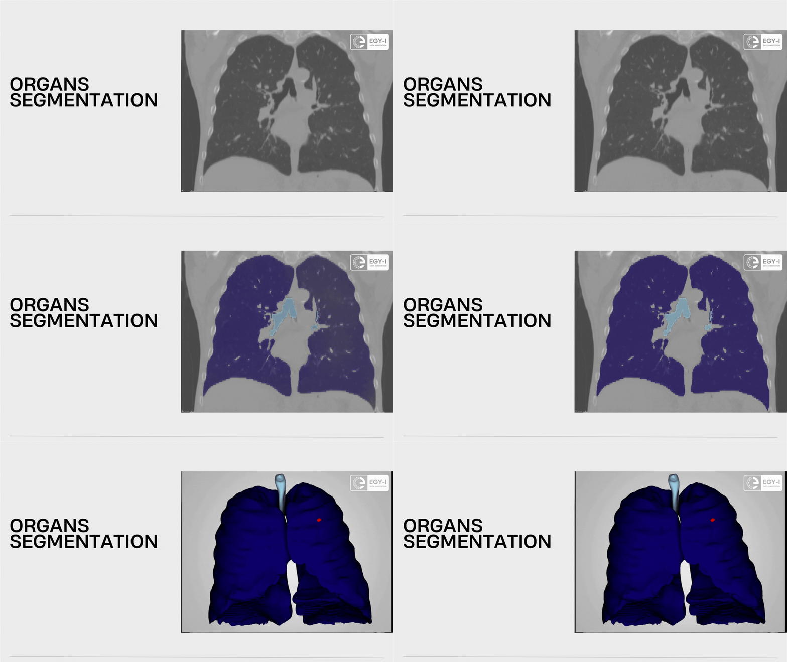
- Organ Segmentation: Precise delineation of organs like the brain, lungs, or liver.
- Pathology Labeling: Categorizing abnormalities such as hemorrhages, infarcts, or calcifications.
- Artifact Detection: Identifying motion artifacts or other image distortions to enhance AI model training

Benefits of AI in CT and MRI DiagnosticsIntegrating AI into CT and MRI workflows delivers numerous benefits:
- Improved Accuracy: AI reduces diagnostic errors by identifying subtle abnormalities and inconsistencies in images.
- Faster Turnaround Times: AI’s ability to analyze images in seconds speeds up the diagnostic process, critical in emergency settings.
- Enhanced Patient Outcomes: Early and accurate detection of conditions leads to better treatment plans and improved prognoses.
- Reduced Radiologist Workload: AI automates repetitive tasks, allowing radiologists to focus on complex cases.
- Cost Efficiency: AI-driven workflows reduce the need for repeated imaging and unnecessary procedures.
How EGY-I Supports AI Development in CT and MRI;
At EGY-I, we provide tailored medical annotation solutions for developers working on AI in CT and MRI diagnostics. Our services are designed to meet the unique challenges of medical imaging:
- Expert Annotators: Our team combines clinical expertise with technical precision to deliver accurate annotations.
- Customized Datasets: We provide annotations tailored to your specific AI use case, ensuring optimal results.
- Quality Assurance: Rigorous quality checks ensure every dataset meets the highest standards.
Revolutionizing CT and MRI with AIAs the adoption of AI in medical imaging continues to grow, the potential to transform CT and MRI diagnostics is immense. At EGY-I, we are proud to support this transformation by providing the high-quality datasets that AI developers need to build reliable, cutting-edge solutions.


