How AI Enhances Accuracy in Chest X-Ray Analysis

Chest X-rays are among the most frequently performed imaging studies worldwide, serving as a first-line diagnostic tool for various conditions, from lung infections and chronic diseases to cardiac and skeletal abnormalities. However, interpreting chest X-rays requires high expertise and meticulous analysis due to the complexity of anatomical structures and the subtle nature of many abnormalities.Artificial Intelligence (AI) has emerged as a game-changer in this field, enhancing diagnostic accuracy, streamlining workflows, and supporting radiologists in delivering faster, more precise care. At EGY-I, we play a pivotal role in enabling these advancements by providing the high-quality annotated datasets essential for AI development in chest X-ray diagnostics.
AI Applications in Chest X-Ray Diagnosis
AI in chest X-ray analysis has unlocked transformative capabilities across multiple aspects of diagnosis and care:
1. Automated Detection of AbnormalitiesAI models can identify a wide range of conditions, including:
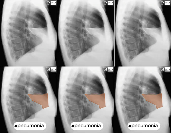
- Pneumonia: Detecting inflamed lung areas with high sensitivity.
- Pulmonary Nodules: Early identification of nodules that could indicate cancer.
- Tuberculosis: Screening for characteristic lung changes with rapid turnaround.
- Pleural Effusion: Recognizing fluid accumulation around the lungs.
- Pneumothorax: Detecting collapsed lung regions in emergency cases.
These systems act as a "second reader," providing radiologists with additional insights and reducing the likelihood of missed diagnoses.
2. Triage and Workflow Optimization: AI-powered systems can prioritize critical cases by flagging chest X-rays that show urgent abnormalities, such as pneumothorax or cardiac enlargement. This triage capability ensures that life-threatening conditions receive immediate attention, improving patient outcomes.
3. Quantitative Analysis: AI can perform precise measurements, such as calculating lung volumes, quantifying lesion sizes, and assessing the progression of conditions like fibrosis or emphysema. These quantitative insights help in treatment planning and monitoring.
4. Enhancing Accessibility: In regions with limited radiology expertise, AI-equipped portable chest X-ray devices can provide reliable diagnostic support, bridging healthcare gaps in underserved areas.
5. Integrating with Decision Support Systems: AI can combine chest X-ray findings with patient histories, lab results, and other clinical data to recommend diagnoses or suggest further tests, enhancing the decision-making process.

Advantages of AI in Chest X-Ray Diagnostics
The integration of AI into chest X-ray analysis delivers numerous benefits for patients, healthcare providers, and medical institutions:
1. Improved Diagnostic Accuracy: AI enhances radiologists' ability to detect subtle abnormalities, reducing diagnostic errors and variability.
2. Faster Turnaround Times: AI-powered systems can analyze chest X-rays in seconds, significantly speeding up diagnosis and enabling quicker clinical decisions.
3. Reduced Workload By automating routine tasks and acting as a second opinion, AI lightens the workload for radiologists, allowing them to focus on complex cases.
4. Early Disease Detection: AI can identify conditions at earlier stages, such as lung cancer or tuberculosis, facilitating timely interventions and better outcomes.
5. Greater Accessibility: In low-resource settings, AI-equipped X-ray devices can provide diagnostic support where radiologists are scarce, democratizing access to quality care.

EGY-I: Empowering AI Innovations
At EGY-I, our specialized chest X-ray annotation services provide the critical datasets needed to train and validate cutting-edge AI models.Key strengths of our services include:
- Expert Team: Our annotators have deep expertise in medical imaging, ensuring annotations align with clinical needs.
- Tailored Solutions: We adapt our annotation processes to meet the unique requirements of each AI project.
- Unmatched Precision: Rigorous quality checks guarantee that our annotations enable AI systems to achieve peak performance.
Whether you’re developing AI for disease detection, triage, or workflow optimization, EGY-I delivers the data you need to succeed.

